Blog Oral Surgery
Manjit singh aged 28 years male, resident of, vill.Balowal Saunkheri Tehsil-Balachaur Dist. Shahid Bhagat Singh Nagar, was brought to the Laungia Dental Hospital. The Patient was brought for treatment only, no medico legal action was needed. Patient was conscious well oriented to time and space, walked normally. He gave a history of road side accident, being hit by a stray cow, while riding his Activa moped. Patient C/o severe pain in the lower jaw, and was not able to chew or close his mouth properly. No history of Head injury and unconsciousness/vomiting.No past history of Epilepsy or Chronic diseases.
On examination : Pulse 80/minute regular, BP 130/80. Signs of Anaemia, Cyanosis nil. Glands, Chest, Abdomen NAD. There was swelling pain and tenderness over the right side mandible with slight mobility of the fractured segments. The X-ray clearly shows the fracture line passing between First & Second Premolars to the body of the Mandible.Hence it is a favorable Mandibular fracture of right side with minimum displacement.
Discussion : This is a ideal case for closed reduction with MMF. In unfavorable cases open reduction is required with plate fixation to keep the fractured segments together.
» Treatment
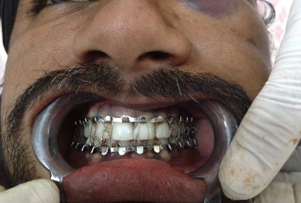
A preventive inj. T.Toxoid and preanesthetic inj. Diazepam was given I.M. With minimum manipulations correct occlusion was obtained.The MMF (Mandibulomaxillary fixation) was achieved by the technique of Arch wiring on Maxilla and Risdon wiring on Mandible.


Postoperative Care : The Patient was educated for postoperative oral hygiene and advised to take liquid/semi liquid nutritious food, strictly to avoid sour, fried, spicy food causing cough reflex/vomiting. Inj. ketanov stat & sos. A course of prophylactic antibiotic Cefadroxil dispersible tabs and analgesic anti inflammatory paracetamol with Ibuprofen liquid was given. A post operative X-ray demonstrated a well reduced fracture line and teeth in proper occlusion.
Maxillary fracture of left side Dated 24-04-2013
A male patient aged about 28 years was brought to Laungia Dental Hospital at 1 P.M. on 24-04-2013 (Wednesday) by relatives from Village Purkhali in Dist. Rupnagar,with a history of accident, involving the patient on cycle, with a unknown person on motorbike.The patient was brought for treatment purpose only, and no medico legal action was required.
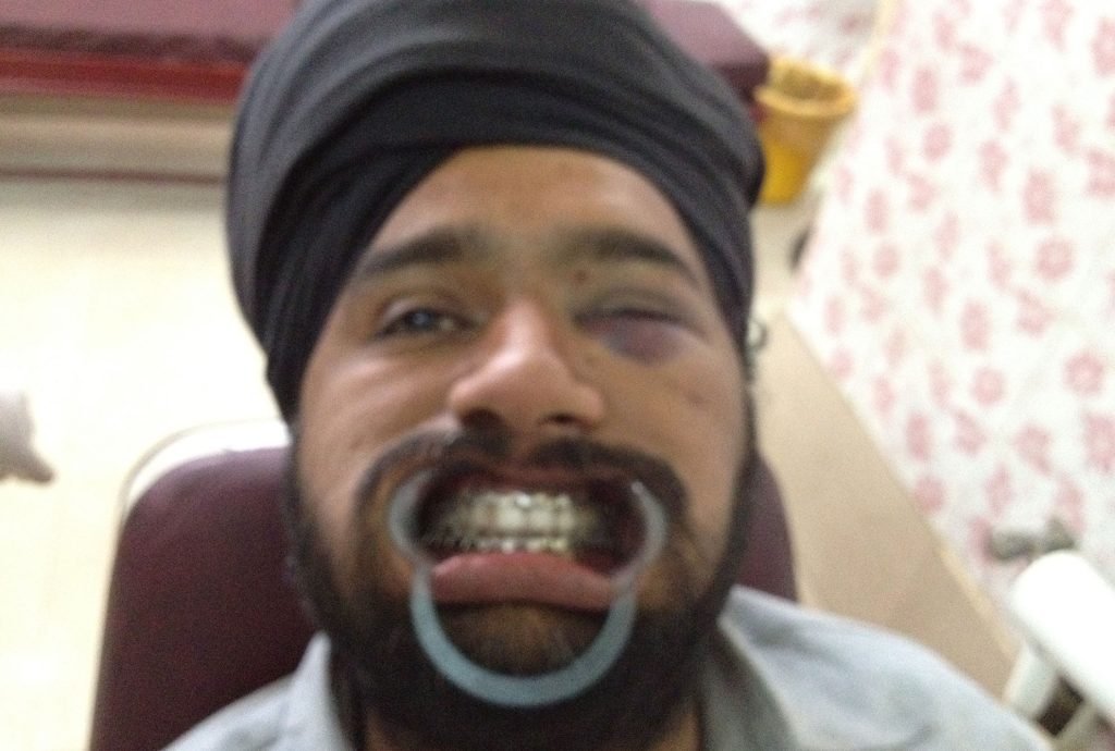
The patient was already examined at some trauma Centre excluding any cranial injury and/or to other vital organs.The patient was conscious and responding to the questions with correct orientation of time & space, C/o pain and swelling on left side face, inability to chew or close his mouth properly. On questioning no history of unconsciousness and/or vomiting was given. On examination Patient had normal B.P. and Pulse rate, with normal movements of all the limbs and no remarkable swelling & tenderness of other body parts. The whole left eye was swollen, with a bluish-black painful, tender swelling at the lower margin of orbit.
There was subconjunctival haemorrhage on left side, but normal pupillary reflexes and movements of eye ball, no diplopia/loss of vision on both sides. There was swelling of whole of the middle third of the left side face. No lacerations or wounds on the face or oral cavity. No bleeding from nose, ears, no active bleeding from mouth. No difficulty in breathing and swallowing. There was significant Mandibulomaxillary Malocclusion. In the A.P. view X-ray, a fracture line at the zygomatic process of left maxilla was seen. This was enough to clinch the diagnosis of fracture of left side maxilla, probably a mixed Lee fort One and Two types of fracture.
DISCUSSION :
The ecchymosis at the infra orbital margin with pain, tenderness & swelling with a fracture line at the zygomatic process of left maxilla in X-ray, points towards Lee fort two type. While no marked flattening of face on Left side, rather a significant mandibulomaxillary malocclusion, later comparatively easy reduction of the occlusion points towards mixed Lee fort type one and two. No neurological sign symptoms and no involvement of any cranial bone in X-ray ruled out Leefort class three.
TREATMENT :
An injection Diazepam I.M. as preanaesthetic and Inj. T.Toxoid preventively was given. Then bilaterally complete mouth block was done with injection lidocaine 2%.
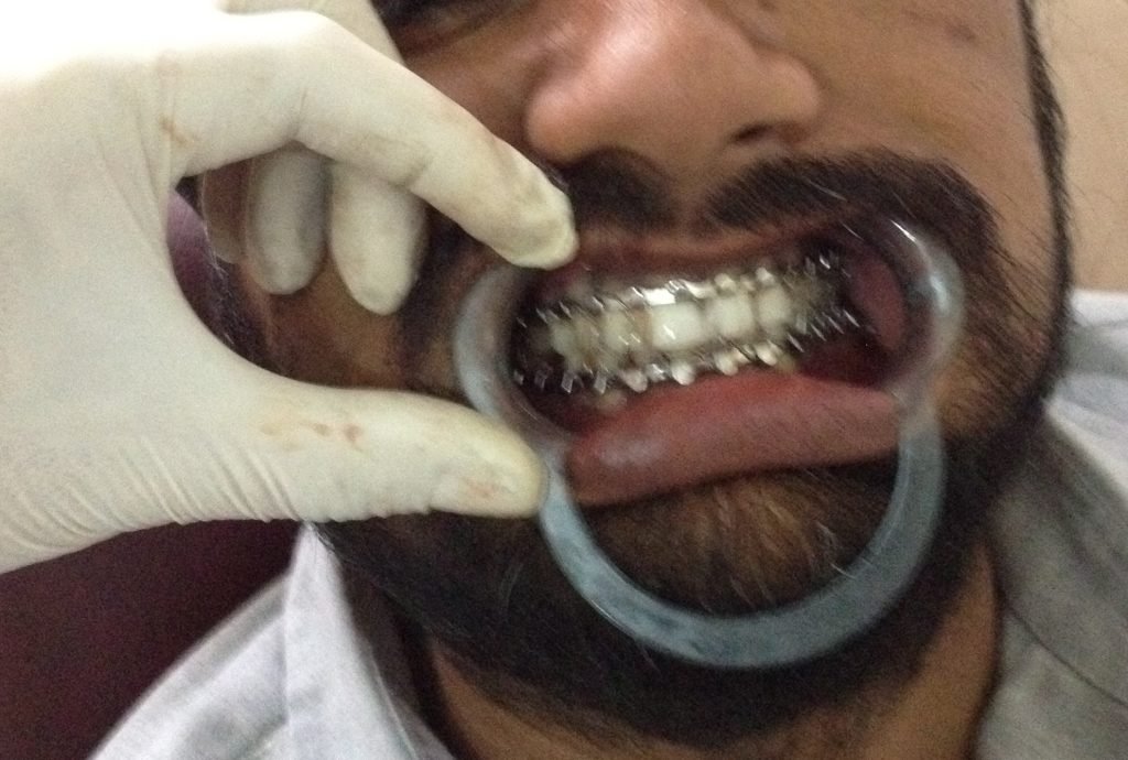
Bilaterally Mandibular Lingual, Posterior superior maxillary, Palatine,Incisive and Infra orbital blocks were done. Then with manipulations the mandibulomaxillary occlusion was restored satisfactorily/completely. Mandibulomaxillary Fixation (MMF) was done with Arch wiring.
POST OPERATIVE :
A prophylactic course of dispersible/Liquid Cefadroxil (antibiotic)and Paracetamol with Ibuprofen (analgesic anti-inflammatory) given for five days with injection Ketanov, stat and sos. Patient advised for liquid/semi liquid diet, no spicy, fried, sour foods, completely avoid cough reflexes, to use antiemetic sos and instructed to get wiring opened in case of any emergency.
In a postoperative P.N.S. view X-ray the fracture line was seen well reduced.
» REPLANTATION OF AN AVULSED TOOTH A PEDODONTIC CASE :
CASE STUDY & MANAGEMENT BY:- Chief Dental Officer Dr.Parminderjit Kaur.
CASE PRESENTED BY:- Dental Officer Dr.Nishant Rajput.
SPECIAL THANKS TO:- Director Dr. Sarbjit Singh Laungia for his Academic inspirations.
DATED:-Presented on 18-04-2014. A case Managed on 28-02-2014.
PATIENT PROFILE:- Arshpal singh a boy aged about 11 years and hence is under pedodontics domain.He is a resident student of an elite educational institution namely Sahibzada Ajit Singh Academy at Rupnagar.
HISTORY:- He had gone to attend a marriage with his father, and there he met an accident, with a fall on floor hitting his mandible. This caused some injury on the chin but avulsion (dislocation) of 1/1 (right upper central incisor) tooth. He and his father immediately and very intelligently saved the avulsed tooth in a clean napkin and first aid was given to his injury. Next day i.e. on 28-02-2014, he came very proudly with his saved tooth in his Institution Uniform, fully confident for restoring back his avulsed tooth.
EXAMINATION:-
(1) There was minor laceration with small bruise on the chin, but no external marks of injury on the upper lip.
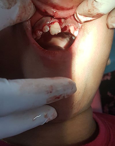
(2) The 1/1 tooth was avulsed and the corresponding socket was filled with clot without any other marked soft tissue injury.
(3) Digital radiographs were taken and the corresponding alveolar socket was found intact and no other injuries detected.
(4) The avulsed tooth was dried up with few debris on it.
(5) Dental occlusion was O.K. with class one relationship. No signs of any other head or significant body injury.
DIAGNOSIS:- It is a case of accidental traumatic avulsion of 1/1 permanent tooth, with minor injuries of the chin. The tooth was not well preserved and brought very late to the Laungia Dental Hospital. The outer vital Cementum layer and the avulsed periodontal ligaments were dried up and dead.
AXIOM:- Avulsion of permanent teeth occurs most often in children of 7 to 9 years old, an age when the relatively resilient alveolar bone provides only minimal resistance to extrusive forces, and the maxillary central incisors are the teeth most commonly affected.
Pics. Showing the effect of avulsion on Periodontal ligament and empty 1/1 socket of the patient with a dressing on the chin injury.

LINE OF TREATMENT:-The aim of the treatment is to fill in the gap and restore the aesthetic and functional part of the avulsed tooth. The first and and most appropriate line of treatment is considered in this case is Replantation of the avulsed tooth. Which involves:-1) Preparation of the Avulsed tooth.2)Replantation and Splinting.
CONSENT:-Patient and the attendants were thoroughly explained about the treatment procedure with a prognosis of quick restoration of structure & functions of the avulsed tooth but also about the gradual resorption of root and finally tooth loss and the expenses involved in it. Patient was also explained about future managements possible and finally a duly written Consent was obtained.
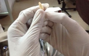
1) Preparation of the tooth:-The carefully brought tooth by patient was unwrapped from the napkin and immediately immersed in to the normal saline solution. All the debris were removed from the tooth. With the avulsion of tooth all the apical vessels & Nerves are lost permanently. Hence an in Vitro One sitting root canal treatment of the tooth was done. The tooth root canal was opened satisfactorily and all the dead vessels & nerves were evacuated completely. The root canal was thoroughly irrigated with Hydrogen peroxide then rinsed with normal saline and antibiotic solution. Special care was taken to avoid any contact of Hydrogen peroxide tot the tooth outer surface. Obturation was done with complete filling of the root canal with Gutta percha. As the Apex was in good shape retrograde filling was not considered. Post obturation was done with Composite material and exposed to light curing. Finally the prepared tooth was cleaned with Normal Saline and thoroughly bathed with antibiotic solution.
Pics. Showing Examination of the Avulsed tooth.
REPLANTATION:- pt had already received a Tetanus Toxoid booster dose. The infra orbital on right side and incisive block anaesthesia was given with Inj. 2% lidocain. The socket of the avulsed tooth was cleaned properly. The previously prepared and treated avulsed tooth was implanted snugly in to the prepared socket with correct Anatomical & Occlusal position. Finally it was firmly splint fixed by wiring to the adjacent left upper central incisor tooth 2/1. Minor wound of the chin was also attended.
Pics.
1) Showing Replantation.
2) Finally splint Fixed avulsed tooth by wiring to adjacent tooth.

FOLLOW UP & AFTER CARE:- Patient was given antibiotic Cephadroxil 250 mg. twice daily for one week with analgesic Ibuprofen 200 mg. twice daily for five days. patient was advised for proper oral hygiene using soft brush and Clohexidrine mouth wash. Patient was advised to come for fortnightly check ups, and given an appointment for removing the splint wiring after two months.
FROM DIRECTORS PEN:- With great satisfaction about Academic Profile of Laungia Dental Hospital. I am making an academic discussion as follows.
DISCUSSION:-
(1) This case reflects a positive impact of Dental Health Education and Awareness about the Modern Dental Technology. We succeeded as far as inspiring & educating the patient & his guardians about Replantation of avulsed tooth.But the tooth was not well preserved and brought in time.
(2) The tooth must be preserved in some Bio compatible & Isotonic Solution to blood & Tissue Fluids, like:-Normal saline/Fresh Cow’s milk/Own Saliva etc. at body temperature, and must reach a Dental Speciality with in one hour. This is to keep the outer vital layer of the tooth namely cementum and the avulsed periodontal ligaments attached to it healthy and living. A well preserved tooth with living vital outer layer after replantation can again regrow the avulsed periodontal ligaments from the blood supply of the socket.This gives the natural resilient fixation of the tooth to the socket, while it results in firm bony ankilosed fixation in case of improperly preserved & brought tooth with dead outer vital layer. Of course the inner layers (Dentine & Pulp) are always dead in an avulsed tooth due to loss of Apical vessels & nerves. Hence the need of R.C.T. in Vitro for all cases of replantation.
(3) Now the most important part of the Discussion is, If in this particular case tooth was not saved and lost. What could be the line of treatment? This patient is aged about 11 to 12 years old and is discussed from Pedodontics considerations. In this age group the both upper and lower Premolars, and second Molars, may not be well erupted and surely their roots are not completed (with open apex). So there is pretty need for movements of the Dentition to accommodate their own growth and that of the Mandible & Maxilla. So such patients if they don’t save the avulsed tooth can not enjoy a fixed prosthodontic Bridge or Dental Implant. Then the only alternative left is RPD ( removable partial denture) which though not very pleasant but would be a must to fill the gap.
(4) Lastly I take the privilege of discussing the Medico-Legal aspect. There was no significant mark of external injury on the corresponding parts of the upper lip in relation to the avulsed tooth. There was little laceration and bruise of the chin but no injury to lower teeth. The Mechanism involved in avulsion of the upper incisor tooth was a transmission of Force to the Avulsed tooth by a Counter coup injury caused by injury to the chin, due to a mobile Mandible and Fixed maxilla by courtesy the resilient soft tooth socket in this age group.
